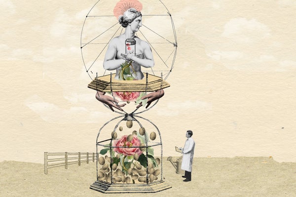
Women live far longer than they used to in the U.S., and five years longer than the average U.S. man. As a result, many believe that they’re the healthier sex. But U.S. women actually spend more of their late lives in poorer health or frailty. In other words, their healthspan falls far short of their lifespan. And a lot of that comes down to their ovaries.
Ovaries, the female gonads, play a pivotal role not only in reproduction but also in overall health. They are responsible for producing eggs and endocrine hormones, including estrogen and progesterone, which are critical for maintaining the menstrual cycle. The effect of these hormones on overall health is apparent at the age of menopause, when ovaries stop working. Postmenopausal women are at heightened risk for conditions such as osteoporosis and heart disease.
Even though lifespans for women have increased to 80 in the U.S., the age of menopause has stayed more or less fixed at 52 years on average, which means women live for decades with a compromised endocrine system.
Because women spend more of their late lives in poorer health or frailty, the concept of healthspan must encompass the effect of the reproductive organs on women’s health as they age. If the ultimate goal is to align healthspan with lifespan—to ensure that women stay healthy throughout their lives—scientists will have to find a way to extend the benefits of healthy ovaries to women after they have passed menopause.
A fast-aging organ
Aging is associated with a general deterioration of tissue function that underlies numerous chronic conditions and illnesses. The female reproductive system is unique in that it is one of the first in the human body to show overt signs of aging. Fertility begins to decline in women in their mid-30s, and reproductive function ceases completely at menopause. Reproductive aging is occurring in females in the prime of their life.
The ovary is the female gonad, which houses egg cells. Women are born with all the eggs they will ever have, and each egg is surrounded by supporting cells within a follicle—the functional unit of the ovary. A major hallmark of ovarian aging is the loss of egg quantity.
Primordial follicles, formed in the womb, make up the ovarian reserve, which is finite and nonrenewable. The ovarian reserve naturally declines across a female’s reproductive lifespan. Females have about a million primordial follicles at birth and a few hundred thousand at puberty. By menopause, only about a thousand primordial follicles remain. Many factors can accelerate the natural age-dependent trajectory of follicle loss—genetic conditions such as X-chromosome abnormalities; smoking and environmental contaminant exposures; and medically induced treatments such as chemotherapy and radiation.
As the number of egg cells drops, the quality of the eggs that remain also decreases. For example, the incidence of chromosomal abnormalities, or aneuploidy, in eggs from women of advanced reproductive age increases dramatically. These abnormalities manifest when the cell undergoes final stages of maturation prior to ovulation. A recognized example of this phenomenon is trisomy 21, or an extra copy of chromosome 21, which results in Down syndrome. The likelihood of having a child with Down syndrome increases significantly in women as they age.
Although a multitude of mechanisms contribute to the decrease in egg quality with age, many are related to the fact that the egg is an extremely long-lived cell. Eggs are formed in utero during fetal development, but are not ovulated until decades later, between puberty and menopause. By then, like a car parked outside through countless winters, the egg is vulnerable to accumulation of damage.
The precise relationship between egg quantity and quality is not completely understood, but it is possible that they are intertwined. As follicles grow, they produce and secrete signals that positively influence the growth and development of nearby follicles. As the number of follicles declines with age, so do the levels of these important signals.
Rising risk
The decrease in both egg quantity and quality together contribute to adverse clinical outcomes. For example, women of advanced reproductive age are at a higher risk of infertility and may need to rely on medically assisted reproduction to conceive. Furthermore, even if women in this age group can conceive on their own, they are at higher risk of spontaneous abortion or are more likely to have twins. They are also at increased risk of complications during pregnancy or having offspring with congenital defects. As more women globally delay childbearing, the negative effect of aging on fertility is having tangible societal consequences, such as more difficulty achieving and maintaining pregnancies and greater prevalence of birth defects. The age at first time birth of women in the U.S. has increased from 25.6 years in 2011 to 27 years in 2024, and this upward trajectory is not only continuing but is a record high.
Although reproductive aging occurs in males and can have measurable impacts on the health of sperm and epigenetic effects on the next generation, the male reproductive system ages more slowly than that of females. For example, the likelihood of a woman being able to conceive without medically assisted reproduction after her mid-40s is exceedingly rare. In contrast, men have been reported to father children into their 90s.
Infertility in general can have profound effects on quality of life and well-being. Women are at a distinct disadvantage to men as they face the biological realities of a reproductive system that ages at what is often considered to be the prime of life. As a physician in obstetrics and gynecology once noted, the ovaries reach their peak potential long before their first use.
The impact of female reproductive aging is much broader than fertility. As the follicles containing eggs grow and develop in the ovary, they produce and secrete estrogen and several other hormones. Estrogen is a steroid hormone that regulates reproductive organs and sexual health. It also controls multiple downstream organs, including the brain, heart, skin, musculoskeletal system and immune system. The loss of follicles with age causes estrogen levels to drop, which can have a plethora of effects on overall health. For example, low estrogen levels can result in bone loss and osteoporosis, as well as increased risk of atherosclerosis and cardiovascular disease.
Medical interventions and health advances are fundamentally changing the way women live. Although the age at which women reach menopause has stayed constant for several centuries, lifespan has been extended significantly. Only 100 years ago, the average lifespan for women in the U.S. was approximately 55 years; it is now 80 years. (Average life expectancy of men in the U.S. is 74.8 years.) This means that women are living nearly three decades with the altered endocrine environment and negative general health consequences of reproductive aging, even though menopause is a part of normal physiology and not a disease.
There appears to be a relationship between menopause and life expectancy: premature menopause is associated with shorter life expectancy and later menopause with longer life expectancy. The ability to extend reproductive function beyond midlife would better align childbearing years and overall healthspan with current longevity.

Katy LeMay
Extending ovarian function
Age-related infertility is primarily rooted in defects that occur at the level of the egg, instead of other organs such as the uterus, according to large-scale data from medically assisted reproduction and specifically in vitro fertilization. Conceiving and giving birth to a child carries a strong maternal age effect, meaning that the risk of complications increases when eggs come from an older woman.
The biological age of the egg is critical in reproductive outcomes. If a woman in her forties undergoes medically assisted reproduction with her own eggs to conceive, it is highly unlikely that she will take home a baby. However, if a woman instead uses donor eggs from a young healthy woman (typically in her 20s) to conceive, the chances are far higher. These observations support the use of planned fertility preservation, or egg freezing, at a younger age with the intent of using them later. But this approach only addresses fertility, not the endocrine function of the ovary. It does not eliminate the inherent pregnancy risks in older individuals and is not a guarantee of future parenthood.
Reproductive longevity can also be extended with hormone replacement therapy (HRT), designed primarily to manage symptoms of the menopausal transition. HRT involves administration of supplemental estrogen and sometimes progesterone, which the ovary no longer produces at sufficient quantities. HRT reduces common perimenopausal symptoms, such as hot flushes, vaginal dryness, mood swings and sleep disturbances.
HRT, however, is a weak substitute for a pair of healthy ovaries. Estrogen replacement does not address fertility. It is generally not prescribed for long-term use due to potential increased risk of breast cancer and cardiovascular disease. And it leaves out the other ovarian steroid hormones, including androgens, and myriad nonsteroidal hormones, such as activin, inhibin, follistatin and anti-Müllerian hormone, as well as growth factors. Furthermore, it is not clear whether the steady hormone levels achieved during HRT are an adequate substitute for the dynamically fluctuating levels of hormones characteristic of natural ovarian cycles.
Emerging strategies
Because of these limitations, we clearly need better, more holistic therapeutic interventions that address both fertility and endocrine function. One potential strategy is to delay reproductive aging by preserving ovarian tissue. The idea is that women would remove and freeze a portion of their ovaries at a young age. The ovarian tissue that is left in the body can compensate for what was removed. We know this because women who have one of their ovaries removed do not undergo menopause appreciably earlier than women with both ovaries intact. As women approach perimenopause, they would then thaw their cryopreserved tissue and have it transplanted back. This method was originally developed for women undergoing fertility-threatening treatments for cancer and other diseases. Hundreds of babies have been born as a result of this procedure. The transplanted ovarian tissue can sustain endocrine function for several years, and ovarian function can be maintained for longer periods with repeated transplants.
Although potentially an attractive option for extending reproductive longevity, this strategy requires an invasive surgical procedure. Ovarian tissue cryopreservation is an acceptable fertility preservation technique for cancer patients, according to guidelines of the American Society for Reproductive Medicine, but using the procedure to extend reproductive longevity in healthy individuals requires further research.
Another attractive target for sustaining ovarian function is the tissue microenvironment—the “nest” in which eggs develop. Several years ago, my laboratory discovered the importance of the microenvironment in which the follicle develops and how it changes with age. When we isolate mouse follicles and eggs from the ovary for research purposes, we poke the ovaries with needles under a microscope. We found that it was more difficult to isolate follicles from ovaries from old mice compared to young ones because the tissue was physically tougher.
This observation reminded us of fibrosis, a biological process characterized by accumulation of collagen proteins that form a matrix or scaffold of tissues. Fibrosis is associated with inflammation, a natural reaction in response to infection or tissue damage. Fibrosis and inflammation are hallmarks of several aging tissues, including the lung, kidney and heart; if not properly resolved, tissue dysfunction ensues. When we looked into this phenomenon, we found that aging mouse and human ovaries tend to stiffen in the same way.
These changes in the aging ovary have significant implications for aspects of ovarian function, including follicle growth, egg quality and ovulation. They may also have implications for how ovarian cancer develops, since cancer cells prefer to colonize stiff, collagen-rich tissues. Several research teams, including my own, have found in preclinical studies that drugs with anti-fibrotic properties can improve reproductive function and longevity. By administering drugs that reduce fibrosis, we can maintain the quality of the ovarian microenvironment, or nest, and prevent key age-related changes in ovarian function both at the level of the egg and endocrine function. These findings are laying the foundation for early-phase clinical trials in women, hopefully in the next few years.
Strategies to improve ovarian function in women beyond middle age raises many questions. Would women want to extend their healthspan if a potential consequence was also extending their childbearing years? Will women who choose to have children in their 50s or 60s and beyond have to deal with age-associated pregnancy risks, or will improved healthspan negate such concerns? How far can we push reproductive longevity? Because women are born with a finite ovarian reserve, there presumably will be a limit to how long ovarian function can be extended, but what is that end point? Could sustained physiological levels of ovarian hormones eventually have negative health consequences? Since the prospects for extending reproductive longevity are likely to occur in our lifetime, we must be prepared to address these safety and ethical considerations.
A priority on health
As drugs are developed that target the mechanisms of aging to prevent age-related conditions and diseases, these therapeutics may not have the same efficacy in both sexes because of unique underlying biology.
The biology of males and females, driven in part by unique sex hormones and X and Y chromosomes, are different at the level of molecular and cellular mechanics. As a result, the same organs may age differently in males and females. This translates into sex differences in age-related diseases. For example, women have a higher incidence and increased death rates compared to men from Alzheimer’s disease and dementia, hypertension and other types of heart disease, chronic obstructive pulmonary disease and kidney disease. Women also have more vision impairments, worse respiratory function and reduced muscle strength and physical performance.
Because research findings on one sex are not typically representative of all, preclinical studies aimed at extending healthspan must include both male and female model organisms. Sex differences may also impact the way drugs work, a fact that will be imperative to acknowledge once compounds enter clinical trials. For example, females are more likely than males to experience adverse drug reactions.
Reframing the discussion of healthspan to include reproductive and women’s health raises new questions and offers new opportunities for growth and discovery. Can signals released by the aging ovary set off a cascade of aging across tissues in women? Are the mechanisms of reproductive aging conserved across tissues? Could reproductive health serve as a biomarker—a canary in the coal mine—for overall health?
How women’s reproductive organs age encompasses nearly all of the biological characteristics of aging overall. We clearly have a lot to learn from ovaries.
Explore the emerging science of healthspan in other stories in this special report.


